Camptochlamys alaskensis Waller & Marincovich, 1992
WALLER, T. R. & L. MARINCOVICH JR. 1992. New species of Camptochlamys and Chlamys (Mollusca: Bivalvia: Pectinidae) from near the Cretaceous/Tertiary boundary at Ocean Point, North Slope, Alaska. Journal of Paleontology, 66 (2): 215-227. [p. 220, figs. 4.1-4.16]
1992 Camptochlamys alaskensis Waller & Marincovich, 1992
T. R. Waller & L. Marincovich Jr., 1992, figure 4.
|
«Etymology. — After the state of Alaska.
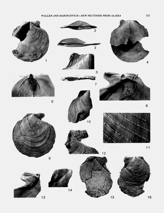
Material. — Holotype, USNM 455515 (Figure 4.1-4.6), a pair of matching valves, the right missing most of its posterior half, the left valve missing its ventral half. Height = 107 mm, length = 102 mm. Paratypes (USNM 455516-455524, 456303-456315) include four articulated specimens variously fragmented, one complete right valve, and about 100 fragments. Many of the fragments are anterodorsal or posterodorsal pieces that allowed tabulation of measurements of auricular shape. Type locality. — Locality M-8120 (Figures 1, 2), in Ocean Point beds, Prince Creek Formation, exposed on bluffs 3 km upstream from Ocean Point, Alaska, 45 km south of the Arctic Ocean. Diagnosis. — Camptochlamys of large size (greater than 100 mm in height), with radial costae present on disk from near umbones to a shell height of at least 25 mm and commonly to more than 60 mm, then fading distally; free margin of posterior auricle forming an obtuse angle (120-133°) with hingeline. Description. — Shell outline: Shell large, height and length about equal, commonly 90-100 mm with maximum reconstructed size about 125 mm. Disk equilateral, anterior half-length usually slightly greater than posterior half-length. Major growth axis from beak to ventral margin curved, prosogyrate, somewhat opisthocline. Anterodorsal margin of disk concave, posterodorsal margin straight. Umbonal angle narrow in early growth stages (69-113° on right valve, 110-116° on left), flaring to maximum of 120-130° in large individuals; anterior umbonal angle greater than posterior umbonal angle. Right valve slightly concave or flat; left valve moderately convex, convexity of articulated shells about 10-15 percent of shell height. Disk flanks very low and poorly demarcated from auricles except for right anterior disk flank, which is steep and covered by out-turned inner shell layer. Anterodorsal and posterodorsal disk gapes moderately wide. Right anterior auricle much larger than posterior auricle, with byssal notch initially very deep relative to size of auricle, later becoming relatively less deep (8-11 mm) in large individuals; byssal fasciole broad, slightly depressed, occupying about half the area of auricle; ctenolium with strong teeth, active teeth ceasing to form beyond a distance of 21-27 mm from beak (measured along suture between anterior auricle and disk); dorsal margin of auricle rising dorsally, becoming well elevated above hinge line at its anterior end; distal third of auricle commonly turned medially in large individuals. Left anterior auricle also larger than posterior, its free margin forming an obtuse angle (110-120°) with hingeline, its dorsal margin nearly coincident with dorsal edge of outer ligament area and its distal third turned outward to conform with medial turning of right auricle. Posterior auricles with free margins more obtuse than that of left anterior auricle, forming angle of 120-133° with hingeline; dorsal margin of right posterior auricle somewhat more dorsally projecting than left. Sculpture: Right valve with broadly and irregularly spaced commarginal very shallow growth steps (numbering eight in holotype, spaced 12-15 mm apart just ventral to center of shell) and more closely spaced and regular commarginal lirae (1.5- 3.0 mm apart in center of shell) extending across disk and au- ricles; radial costellae of low relief present on disk beginning at shell height of about 2 mm, increasing by intercalation to pro- duce an irregular alternation in strength, fading distally beyond shell height of between 25 and 85 mm. Left valve with sculpture similar to right but with growth steps less evident, commarginal lirae projecting and flange-like, and radial costellae stronger; radial costellae on some left valves present on disk flanks and proximal parts of auricles as well as on disk. Antimarginal microsculpture present on both valves on at least distal halves of interspaces between commarginal lirae (Figure 4.11), sometimes well developed and extending across entire space between com- marginal flanges. Shell interior: Anterior outer ligament longer than posterior (anterior to posterior ligament length ratios 1.03-1.36, averaging about 1.25), outer-ligament insertions showing evidence of considerable ventral migration of active ligament. Right hinge teeth very weak, with resilial teeth barely raised above hinge plate and dorsal teeth low, each dorsal tooth visible as a sloping broad raised undulation across outer-ligament area; right outer-ligament areas bordered ventrally by sharp but shallow step down to auricular surfaces, this step absent on left valve; ventral edge of resilifer slightly undercut on right valve, deeply undercut on left. Right valve interior somewhat thickened along boundaries between disk and auricles, especially on anterior side, where irregular radial rugosities sometimes form (Figure 4.13). Interior of right auricle with a faint broad radial ridge over boundary between byssal fasciole and dorsal part of auricle. Left valve interior without significant thickening along boundaries between disk and auricles. Adductor scar large, overlapping center of shell but mainly developed to posterior side of center, with its anteroventral boundary on right valve bilobate, separating large central striate-muscle insertion from smaller posterior nonstriate-muscle insertion, area of latter about one-third that of former. Byssal retractor scar present on left valve, indicated by small postero-dorsal extension of left adductor scar. Pallial line well inset from valve margin midventrally (about 39 mm on holotype, where valve height is 113 mm), continuous but somewhat uneven in width, leaving radial tracks on inner surface of outer shell layer; anterodorsal and posterodorsal parts of pallial line, marking insertions of circular pallial muscles, discontinuous and deeply pitted, especially on left valve, and insinuated on anterior (pedal sinus) and posterior (pericardial pocket) sides. Shell microstructure: Shell mainly foliated calcite, with laths arranged in broad irregular arrays outside of pallial line and in small irregular reflective patches inside pallial line; radially lathic outermost shell layer very thin. Aragonite present in muscle scars (myostracum), ligament insertion areas (ligostracum), and as a thin inner shell layer secreted in a restricted region inside the pallial line anteroventral to adductor scar. Prismatic calcitic stage of right valve not preserved but inferred to be no more than 2 mm in height based on start of radial costae. Comparison. — The new species differs from known species of Camptochlamys in having a more flaring umbonal angle and far more obtuse posterior auricles. Compared to the two species of Camptochlamys recognized by Johnson (1984), the new species is intermediate in having radial costae extending further than those of C. obscura but not as far as those of C. clathrata. The new species differs from all known Mclearnia in having radial costae, a thinner shell, greater ventral migration of the ligament system, and greater dorsalward flaring of the dorsal margin of the right anterior auricle relative to that of the posterior auricle. There are no pectinids known from the Paleocene or Eocene that are comparable in morphology or maximum size to the new species. Stratigraphic and geographic range. — The new species is thus far known only from the Ocean Point beds. Within these beds the species is distributed through about 7 m of the section vertically (Figure 2). Ecology and taphonomy. — Camptochlamys alaskensis n. sp. was very likely byssate throughout life, reclining on its flattened right valve on a bottom of firm sediment but with the ability to swim when necessary. Persistent byssal attachment is indicated by five features: 1) a persistent moderately large byssal notch; 2) a strong ctenolium that retained active teeth until late in ontogeny; 3) persistent auricular asymmetry, with the dorsal margin of the right anterior auricle more projecting than that of the posterior; 4) significant ventral migration of the ligament system; and 5) the probable presence of an attachment scar for a left pedal retractor muscle. Another good indicator of a sedentary and probably byssate life habit is the unusual inward turning of the end of the right auricle (and outward turning of its left counterpart). The end of the anterior auricle is one of the contact points with the substrate in byssally attached pectinids (Stanley, 1970; Waller, 1972, 1984), and its upturning may signify persistent pressure or an adaptation to keep the end of the auricle out of the sediment. Swimming ability even among the largest specimens is suggested by both the lightness of the shell and the presence of significant anterodorsal and postero- dorsal shell gapes. The great majority of Pectinidae feed on phytoplankton, which is removed from water pumped through the filibranchiate ctenidia by strong ciliary action. Some pectinids, however, also feed on organic detritus resuspended as a result of valve clapping. The latter has been documented in living Placopecten magellanicus (Gmelin, 1791), a Western Atlantic cold-temperature species with a flattened right valve (Shumway et al., 1987). It is also a likely mode of feeding for the Antarctic scallop Adamussium colbecki (Smith, 1902), which lives in an area of highly seasonal productivity (Berkman et al., 1991). The shells of Camptochlamys alaskensis n. sp. have an epifauna of low diversity. Chalky, calcareous polychaete worm tubes occur on many of the specimens. On right valves these tubes are limited to the distal parts of the disk and to the posterior auricular region. Their growth is mainly linear following an early coiled stage, and where they intersected the growing shell margin, the margin formed a small deflection. On the left valves the worm tubes are less localized and have only the coiled stages; no marginal deflections were observed. On some specimens (Figure 4.9) only a raised flattened sinuous track on the shell surface is present. Presumably this is all that remains after destruction of the aragonitic worm tube by weathering and differential etching of the calcitic shell surface. Small colonies of a membranimorph cheilostome bryozoan are present near the distal edge of the right-valve fragment (USNM 455522) that is the basis for the estimate of maximum size given in the description. The poor state of preservation of these cheilostomes allows no independent age determination other than the broad range of Aptian to Recent (A. Cheetham, personal commun.). One left-valve fragment (USNM 455523) has a small (2 mm) borehole with a beveled shape like that known to be produced by predatory naticid gastropods. This borehole was almost certainly made by Amauropsis sp., an undescribed new species that is very common in the Ocean Point fauna. The left valve of one articulated specimen (USNM 455524) and some fragments bear small (less than 5 mm) specimens of an unidentified cementing bivalve that is possibly a new species of Placunopsis. A few specimens have etched pits and grooves suggestive of clionid sponge borings. Lastly, there is abundant evidence of minor marginal fracture and repair in many of the specimens; a few have large repaired fractures that may indicate unsuccessful predation by crabs or fish. Three of the articulated specimens still contain a sediment filling that is of finer grain than the surrounding sediment. These specimens were also severely crushed, suggesting that they were buried alive, finer sediment then infiltrating the valves after the decay of soft parts. Diagenetic compaction then probably operated preferentially on these specimens because their sedimentary filling was less competent to withstand stress than the surrounding sediment. Observations in the field established that the articulated specimens were in living position, with the right valve downward. The state of preservation of the shells is unusual in that the shell surfaces are finely etched, bringing out in relief the fine crystalline grain of the calcitic shell layers. In some cases there are minute perforations and iron-oxide lined cavities following the crystalline fabric of the shell. These were possibly caused by modem fungi.» THOMAS RICHARD WALLER & LOUIE MARINCOVICH JR., 1992
|